Mapping sugar
A new technique makes it possible to image the spatial structure of polysaccharides using a scanning tunnelling microscope
There is a new perspective on sugar. A team of scientists from the Max Planck Institute for Solid State Research and the Max Planck Institute of Colloids and Interfaces has used a scanning tunnelling microscope to image how individual polysaccharide molecules are folded for the first time. The researchers are thus making it possible to investigate the relationship between the spatial structure of polysaccharides such as those found on pathogens and their biological effect.

Function follows form. To a certain extent, the opposite of this guideline mainly followed by Bauhaus designers applies in biology: the form of a biomolecule determines its function. This has long been known about enzymes and other proteins that fulfil their tasks only once they are properly folded. “But until now, it was unclear whether polysaccharides also fold like proteins”, says Peter Seeberger, Director at the Max Planck Institute of Colloids and Interfaces in Potsdam and professor at the Freie Universität Berlin. “We have now found a way to investigate this question”. And indeed: Some polysaccharides such as cellulose do fold, while others such as mannose on the surface of corona viruses or HIV do not.
The sugar specialists led by Peter Seeberger owe their findings on the conformation of polysaccharides to a new method developed by a team led by Klaus Kern, Director at the Max Planck Institute for Solid State Research in Stuttgart. “Scanning tunnelling microscopy allows us to image individual sugar molecules”, says Kern. “Other methods reveal only the average structure of the sample molecules”. But an average structure is of little use, especially for investigations into how the biological function of sugars depends on their form.
After a soft landing, the profiling begins

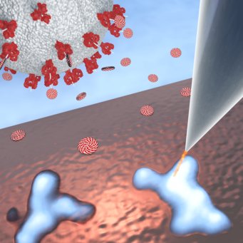
When profiling sugar molecules, they must first be deposited on a surface without becoming deformed. Stephan Rauschenbach, now professor at Oxford University, has developed a method at the Max Planck Institute in Stuttgart that makes this possible. For this purpose, the researchers use electrospray ionization to convert the polysaccharides to the gaseous state. They atomise a solution of the respective substance with a fine nozzle while simultaneously ionizing the molecules with a strong electric field. Using a weak electric field, they bring the charged molecules to a soft landing on a copper surface. Then they begin with the actual profiling. Using the tip of a scanning tunnelling microscope, they scan a sugar molecule on the surface. To keep the particle still during this process, they cool the copper surface to almost −270°C. This gives them a profile drawing of the molecule – quite similar to how a pencil can be used to trace the grain of wood.
With the portraits of the sugars in profile alone, the question of the biologically relevant conformations cannot yet be definitively answered. After all, the polysaccharides could take on a different form as a gas than in dissolved or crystalline form. In close cooperation, the teams from Stuttgart and Potsdam are investigating this question as well as how the surface might affect the structure. “However, the images of the individual molecules provide us with initial useful information that helps us to better understand the relationships between the structure and properties of sugars”, says Seeberger.
PH


