The geometry of cancer cells
Malignant and healthy cells display characteristic fractal patterns, which can be used to tell them apart

The concept of fractals was introduced less than 40 years ago; however, fractal structures have existed from time immemorial. The leaves of a fern, Romanesco broccoli and coastlines all follow a pattern in which geometrical and topographical features are repeated, when they are increasingly magnified. Outgrowth and protrusions on the cell surface also display such “self-similar” patterns.
The team working with Joachim Spatz, Director at the Max Planck Institute for Intelligent Systems in Stuttgart and Professor at the University of Heidelberg, has now discovered that tumour cells and healthy cells can be identified using fractal geometry.
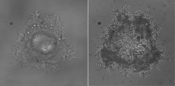
The scientists magnified the contours of human pancreas tumour cells and analysed their physical irregularities. They then calculated the fractal dimensionality, a measure of the statistical distribution of the irregularities, of the cell contours. Cancer cells display a greater degree of fractality than healthy cells, since the chaotic growth of tumours causes very irregular convexities of varying size on the cell surface. However, not only did the fractal dimension indicate for the researchers the presence of a tumour cell, it could also be used to determine with 97 per cent accuracy to which of two different lines of malignant pancreas tumour cells it belonged. “This is a much more accurate and faster method for determining cancer cell types than the established procedure,” says Joachim Spatz.
Determining cancer cell types is still an uncertain and time-consuming task

To date, cancer cells and their primary site of origin in the body were identified in extracted biopsy samples that were stained using specific antibodies and biomarkers. However, there are drawbacks to this method of staining: It requires numerous individual steps with expensive antibodies, rendering it costly and time-consuming. Moreover, the stains predominantly used make it more difficult to distinguish the tiny differences in cells. As a consequence, cancer is only correctly diagnosed in 85 per cent of samples.
Using fractal geometry, Joachim Spatz’ team is able to identify cancer cells more reliably and much faster. With this method, cells can be studied under a microscope without requiring special preparation. In a reflection interference contrast microscope (RICM), the team is able to study the details of the cell contours. Whereas a conventional bright-field microscope illuminates the sample from below, the microscope used by the Stuttgart-based scientists measures the reflection of light on the cell surface. This differs according to whether the light falls directly on a cell or first hits an aqueous cell culture medium and then a cell. The reflected light permits the study of even minute structures on the cell surface.
“Analysis of the fractal geometry of cell surfaces promises great potential for clinical diagnostics,” says Spatz. The scientists are now investigating possible clinical applications for their method. They are studying different malignant cell lines and primary cells, i.e., cells that are taken directly from human organs and, in contrast to tumour cell lines, have a limited culture lifespan. “Our next step is to seek cooperation with clinics, in order to test the method directly on relevant tissue samples,” Joachim Spatz explains.
NG/KH/PH


