An ultrasonic projector for medicine
A chip-based technology that modulates intensive sound pressure profiles with high resolution opens up new possibilities for ultrasound therapy
A chip-based technology that generates sound profiles with high resolution and intensity could create new options for ultrasound therapy, which would become more effective and easier. A team of researchers led by Peer Fischer from the Max Planck Institute for Intelligent Systems and the University of Stuttgart has developed a projector that flexibly modulates three-dimensional ultrasound fields with comparatively little technical effort. Dynamic sound pressure profiles can thus be generated with higher resolution and sound pressure than the current technology allows. It should soon be easier to tailor ultrasound profiles to individual patients. New medical applications for ultrasound may even emerge.

Ultrasound is widely used as a diagnostic tool in both medicine and materials science. It can also be used therapeutically. In the US, for example, tumours of the uterus and prostate are treated with high-power ultrasound. The ultrasound destroys the cancer cells by specific heating of the diseased tissue. Researchers worldwide are using ultrasound to combat tumours and other pathological changes in the brain. “In order to avoid damaging healthy tissue, the sound pressure profile must be precisely shaped”, explains Peer Fischer, Research Group Leader at the Max Planck Institute for Intelligent Systems and professor at the University of Stuttgart. Tailoring an intensive ultrasound field to diseased tissue is somewhat more difficult in the brain. This is because the skullcap distorts the sound wave. The Spatial Ultrasound Modulator (SUM) developed by researchers in Fischer’s group should help to remedy this situation and make ultrasound treatment more effective and easier in other cases. It allows the three-dimensional shape of even very intense ultrasound waves to be varied with high resolution – and with less technical effort than is currently required to modulate ultrasound profiles.
High intensity sound pressure profiles with 10,000 pixels
Conventional methods vary sound fields with several individual sound sources, the waves of which can be superimposed and shifted against each other. However, because the individual sound sources cannot be miniaturized at will, the resolution of these sound pressure profiles is limited to 1000 pixels. The sound transmitters are then so small that the sound pressure is sufficient for diagnostic but not therapeutic purposes. With the new technology, the researchers first generate an ultrasonic wave and then modulate its sound pressure profile independently, essentially killing two birds with one stone. “In this way, we can use much more powerful ultrasonic transducers”, explains postdoctoral fellow Kai Melde, who is part of the team that developed the SUM. “Thanks to a chip with 10,000 pixels that modulates the ultrasonic wave, we can generate a much finer-resolved profile”.

“In order to modulate the sound pressure profile, we take advantage of the different acoustic properties of water and air”, says Zhichao Ma, a post-doctoral fellow in Fischer’s group, who was instrumental in developing the new SUM technology: “While an ultrasonic wave passes through a liquid unhindered, it is completely reflected by air bubbles”. The research team from Stuttgart thus constructed a chip the size of a thumbnail on which they can produce hydrogen bubbles by electrolysis (i.e. the splitting of water into oxygen and hydrogen with electricity) on 10,000 electrodes in a thin water film. The electrodes each have an edge length of less than a tenth of a millimetre and can be controlled individually.
A picture show with ultrasound

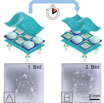
If you send an ultrasonic wave through the chip with a transducer (a kind of miniature loudspeaker), it passes through the chip unhindered. But as soon as the sound wave hits the water with the hydrogen bubbles, it continues to travel only through the liquid. Like a mask, this creates a sound pressure profile with cut-outs at the points where the air bubbles are located. To form a different sound profile, the researchers first wipe the hydrogen bubbles away from the chip and then generate gas bubbles in a new pattern.
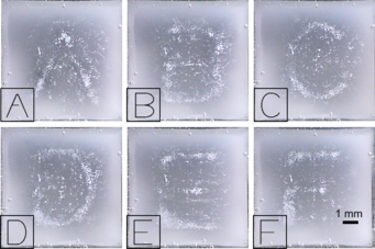
The researchers demonstrated how precisely and variably the new projector for ultrasound works by writing the alphabet in a kind of picture show of sound pressure profiles. To make the letters visible, they caught micro-particles in the various sound pressure profiles. Depending on the sound pattern, the particles arranged themselves into the individual letters.
Organoid models for drug testing
For similar images, the scientists collaborating with Peer Fischer, Kai Melde, and Zhichao Ma previously arranged micro-particles with sound pressure profiles, which they modelled using a slightly different technique. They used special plastic stencils to deform the pressure profile of an ultrasonic wave like a hologram and arrange small particles – as well as biological cells in a liquid – into a desired pattern. However, the plastic holograms only provided still images. For each new pattern, they had to make a different plastic template. Using the ultrasound projector, the Stuttgart team is able to generate a new sound profile in about 10 seconds. “With other chips, we could significantly increase the frame rate”, says Kai Melde, who led the hologram development team.
The technique could be used not only for diagnostic and therapeutic purposes but also in biomedical laboratories. For example, to arrange cells into organoid models. “Such organoids enable useful tests of active pharmaceutical ingredients and could therefore at least partially replace animal experiments”, says Fischer.
FM/PH


