Single-celled organisms shed light on neurobiology
Bacteriorhodopsin and channelrhodopsin, which originates from a unicellular green alga, are advancing to become new tools in neurobiology
The discovery of a visual pigment in the cell membrane of an archaebacterium in the early 1970s is owed solely to a researcher’s curiosity: For three years, the scientific community wouldn’t believe Dieter Oesterhelt. Forty years after his pioneering work at the Max Planck Institute of Biochemistry in Martinsried, bacteriorhodopsin and channelrhodopsin, which stems from a single-celled green alga, are gaining ground as new tools in neurobiology
Text: Christina Beck

It was an illustrious group that the Royal Swedish Academy of Sciences invited to the Lilla Frescativägen in Stockholm on December 12, 2013. Optogenetics was the topic on which the Nobel Prize Committee was seeking information. Among the eleven scientists were two from Max Planck institutes, as well as two other researchers who had taken their first steps in this field as young group leaders at Max Planck.
As early as 1979, the discoverer of the DNA code, Francis A. Crick, called it the greatest challenge in the neuro-sciences: to selectively influence a specific cell type in the brain and leave the others unchanged. In his lectures, he speculated that light could serve as a control tool – in the form of locally and temporally restricted impulses of different colors.Thirty years later, this vision is becoming a reality: optogenetics is poised to revolutionize the neurosciences, permitting, as it does, the first ever non-invasive manipulation of neural networks in an organism – from the small nematode Caenorhabditis elegans to the mouse. Someday, perhaps, even in humans. In other words, a method that has the potential to win a Nobel Prize.
The discovery of the first microbial rhodopsin
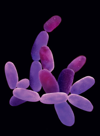
But what has since become a popular tool of neuroscientists had its begin-nings somewhere else entirely: in a small, halophilic archaebacterium, Halobacterium salinarum. Archaebacteria are the “old-timers” of life. Since the early days of evolution, these single-celled organ-isms have persevered in extreme habitats – in salt lakes, for instance, or hot volcanic springs – while bacteria and the eukaryotes were able to develop much more freely.
It was more or less a coincidence that brought biochemist Dieter Oesterhelt into contact with Halobacterium salinarum. But this archaebacterium would eventually become the central research object of his scientific life for the next 40 years. Oesterhelt had done his doc-torate in Feodor Lynen’s lab at the Max Planck Institute for Cell Chemistry (later the Max Planck Institute of Biochemistry) on a metabolic enzyme, fatty acid synthase. “A giant particle,” as he says, “whose structure could be decoded only through electron microscopy.”This explains why the researcher went on a sabbatical in 1969, to San Francisco and Walther Stoeckenius, a renowned expert in the field of elec-tron microscopy. Oesterhelt wanted to learn to use this technology in the lab with him.
Stoeckenius was interested in the membrane of the halobacterium because, at that time, the molecular structure ure of cell membranes was still a subject of controversy. “That was the so-called purple membrane, which is how it was known even then. But it was completely unclear what it was,” says Dieter Oesterhelt.
Allen Blaurock, who was likewise in Stoeckenius’ lab at the time, requested Oesterhelt’s assistance in preparing his samples. Oesterhelt experimented with different organic solvents to elute the lipids from the membrane: “So I extracted the purple membrane with chloroform/methanol – and suddenly I had a yellow extract,” he recalls.
Such a change in absorption across an area of nearly 200 nanometers seemed quite unusual to the young biochemist. But Allen Blaurock dis-missed it – he had worked on frog ret-inas with Maurice Wilkins in London. Their X-ray experiments required them to irradiate the frog retina at a very specific angle. “But if we weren’t careful,” Blaurock told Oesterhelt, “the beam went into the frog’s lovely red eye, and suddenly it turned yellow.”
That was the decisive clue for Diet-er Oesterhelt. He went to the library to retrieve the data on retinal, the light-absorbing pigment in the retina of vertebrates, and then analyzed the purple membrane using mass spectroscopy. No doubt about it: it was retinal. Walther Stoeckenius’ initial reaction, however, was less than euphoric – he said, quite simply: “That can’t be. That doesn’t exist in prokaryotes.”
The reviewer for the journal Nature likely had a similar view. The submitted publication was rejected with a note saying that the experiments were fine, but the analogy with rhodopsin was pretty far-fetched. “It was simply unacceptable to find retinal somewhere other than in an eye,” concludes Oesterhelt. So the first publication on bacteriorhodopsin, as the authors had christened their molecule, appeared in 1971 in the journal Nature New Biology.
Photosynthesis invented not once, but twice
Dieter Oesterhelt returned to Munich and – despite serious doubts on the part of his colleagues there – continued to work with the bacteriorhodopsin: “It seems to me to be quite an unusual thing, and it isn’t there without reason,” he explained to the skeptics. And then the shortage of collaborators was also accompanied by a shortage of equipment. The Max Planck Institute for Cell Chemistry moved out to Martinsried, while Oesterhelt remained in the originally shared labs at the Lud-wig-Maximilians-Universität Institute of Biochemistry. “All I had left was a pH meter, a water bath and a projector,” he says. But this situation proved to be a blessing for the key experiment that followed, “because I simply couldn’t do much more.”
Oesterhelt was firmly convinced that the color change is associated with a function, so he worked on reversing it: “Quite simply, I tried every solvent in the world.” And then here, too, coincidence again played a role. Specifically, if I took ether, added salt, and then went to the window when the Sun was shining, the extract suddenly turned bright yellow; in the dark, the color changed back. That was the desired color change, but what was behind it?
“I simply placed a pH electrode in it,” says Oesterhelt. When the color changed from purple to yellow, protons were released, and when the color changed from yellow to purple, protons were taken up. Accordingly, the extract became acidic in the one case, and alkaline in the other. However, when such a release and uptake of protons takes place in a dense layer, such as a membrane, it would have to create a pumping effect.
The young biochemist imagined a proton pump – that is, a molecule that takes up protons from one direction and releases them in the other. Oesterhelt presented this idea to his dissertation supervisor Feodor Lynen. He said simply: “I don’t believe it, but I certainly hope that you’re right.” If the molecule pumps protons, then it should be possible to measure a pH change in a suspension of bacteria cells.
Dieter Oesterhelt set up his pH meter in the darkroom to see what would happen when he exposed intact cells. He set the pH meter to the highest sensitivity and then turned the light on: “The recorder gave a jerk and the needle shot straight to the upper limit.” In a few days, he had gathered the relevant readings and, with them, the proof that bacteriorhodopsin is, indeed, a light-driven proton pump.
By transporting protons out of the interior of the bacteria cell, a proton concentration gradient is created between inside and outside, and an electrical potential is built up across the membrane. “The process is just like charging a battery,” explains the Max Planck researcher. The energy of the protons flowing back in is used for enzymatic synthesis of ATP (adenosine triphosphate), the energy currency of the cell.
Even more light-switched membrane proteins

This was in line with the chemiosmotic hypothesis proposed by Peter D. Mitchell back in 1961 – which earned him the Nobel Prize in Chemistry in 1978 – for which bacteriorhodopsin thus provided initial evidence. The purple membrane system is, next to the chlorophyll system of green plants, the second light-energy conversion principle of living nature. “In other words, evolution invented the fundamental process of photosynthesis not once, but twice,” says Dieter Oesterhelt.
In the years that followed, bacteriorhodopsin rose to become a model subject in bioenergetics, membrane biology and structural biology. Since the start of the second half of the 1970s, there have been more than a hundred publications on this topic each year. In 1977, Japanese researchers Matsuno-Yagi and Mukohata discovered a further pigment in the purple membrane of Halobacterium salinarum, but it differed from bacteriorhodopsin. It was long speculated that this was a light-activatable sodium pump.
Oesterhelt had since become Director at the Max Planck Institute of Biochemistry in Martinsried, and one of his first doctoral students there was chemist Peter Hegemann. He was originally supposed to isolate this sodium pump, which was called halorhodopsin. But then Janos Lanyi and Brigitte Schobert from the University of California showed that this membrane protein wasn’t pumping sodium ions out of the cell, but rather, it was pumping chloride ions into the cell. This would later take on an entirely new significance.
In 1986, Hegemann began leading a group of his own in Oesterhelt’s department of membrane biochemistry and, in the late 1980s, dedicated himself to a new object of study: the small, unicellular green alga Chlamydomonas reinhardtii. In EMBO Molecular Medicine he and his co-author Georg Nagel later wrote: “When we conducted our experiments more than a decade earlier, we hadn’t expected that the research on the molecular mechanisms of the phototaxis of single-celled algae or the light-driven ion transport in archaebacteria could one day be of interest to the readers of a medical journal.” But the road was rocky, and extremely long.
An alga's red eyespot apparatus was puzzling
As a photosynthetic organism, Chlamydomonas seeks out areas where the light conditions are particularly favorable for photosynthesis. In doing so, it uses its long flagella to propel itself like a little breaststroker. This means that the photosynthesis apparatus of the small green alga doesn’t have to continually be adapted to changing light conditions. Scientists refer to such light-controlled orientation movements as phototaxis. They’ve been known since the 19th century. The light sensor responsible for phototaxis is located in the alga’s red eyespot apparatus.
Kenneth W. Foster, a former stu-dent of Max Delbrück, studied the phototactic movements of Chlamydo-monas as a function of the light’s wave-length in order to obtain information about the light sensor’s properties. Using these so-called action spectra, he postulated – already back in1980 – that the light sensor is a rhodopsin. Furthermore, a few years later, he succeeded in restoring the light-driven movements in “blind” algae by adding retinal. “But the photoreceptor researchers’ field didn’t recognize the significance of these findings at the time,” says Peter Hegemann.
However, the clue that a small, single-celled green alga uses a visual pigment that may be very similar to the one in the human eye aroused his interest. Together with his colleagues, Hegemann labored for ten years to obtain the alga’s photoreceptor in sufficient amounts and appropriate purity for protein chemical analyses, but the tests remained unsuccessful: “The radioactive labels yielded a completely undefined image,” he says. Today, the researchers know that there are ten different rho-dopsins in the eyespot apparatus of Chlamydomonas.
Back then, only the electrophysio-logical measurements produced promising results: the photocurrents published in the journal Nature in 1991 not only showed that the photoreceptor did, in fact, have to be a rhodopsin, but they also revealed something else: unlike in the human eye, the current was quite obviously not transmitted via a chemical signal cascade and thus amplified. Instead, the photoreceptor ap-peared to be coupled directly to an ion channel, as the photocurrents appeared extremely quickly, within just 30 microseconds (millionths of a second) after exposure.
Eight years later, Hegemann, who had since been appointed to a position at the University of Regensburg, made an even more pointed statement in a publication: “We assume that chlamyrhodopsin is part of a rhodopsinion channel complex, or even forms the channel itself.” But the scientific community countered these explanations with similar skepticism as for Oesterhelt’s previous discovery of the first microbial rhodopsin.
It still wasn’t possible to list the key properties of this alleged rhodopsin channel. All electrophysiological derivations were carried out using a suction pipette. The researchers were thus always able to register only the total of the ion currents across a large membrane area, but not the current of a single channel.
A new approach was needed. In 2000, the Japanese Kazusa DNA Research Institute published thousands of newly decoded gene sequences from Chlamydomonas reinhardtii in freely accessible online databases. When looking through these sequences, the researchers in Regensburg discovered two longer sections that were similar to bacterial rhodopsin genes.
Hegemann asked Georg Nagel, then a research group leader at the Frankfurt-based Max Planck Institute for Biophysics in Ernst Bamberg’s department, to test the properties of the proteins encoded by these gene sequences.
Bamberg’s department had already accrued years of experience in the electrophysiological characterization of microbial rhodopsins. In order to document the transport characteristics of bacteriorhodopsin and halorhodopsin under electrically controlled conditions, the researchers had transferred them to the egg cells of claw frogs.
The birth of optogenetics
Nagel and his colleagues now likewise tested the electrical properties of the proteins originating from Chlamydomonas in frog eggs. In June 2002, they presented the findings in the journal Science. It was the long-awaited proof that the algae rhodopsin was indeed the first example of a directly light-driven ion channel, and thus a completely novel membrane protein. After taking up light, channelrhodopsin-1 (ChR1), as the scientists had christened their “baby,” channels protons via the membrane into the cell interior in contrast to the proton pump, bacteriorhodopsin, the ion transport requires no energy.

One year later, the researchers published their findings on the second light-activated ion channel, channel-rhodopsin-2 (ChR2), which, in contrast to channelrhodopsin-1, also channels other positively charged particles, such as sodium ions. They had succeeded in incorporating channelrhodopsin-2 not only in frog eggs, but for the first time, also in human kidney cells.
This publication aroused the interest of Karl Deisseroth and Edward Boyden at Stanford University. The two researchers had already been discussing possibilities for controlling the electrical activity of nerve cells in the intact brain for some time. In March 2004, Deisseroth wrote an e-mail to Georg Nagel and asked whether he could, in the context of a collaboration, get a clone of channelrhodopsin-2. The package from Germany arrived a few weeks later.
Boyden, Deisseroth and Feng Zhang, who had subsequently joined them, considered how to proceed. In the months that followed, the researchers optimized their experimental setup. Using a harmless virus as a gene ferry, they managed to introduce the gene for channelrhodopsin-2 into cultivated hippocampus neurons. Via the promoter, a genetic switch, they were able to control which neuron type produces channelrhodopsin-2. The experiment worked, “incredibly well, actually,” as Deisseroth writes. “Using simple, harmless flashes of light, we were able to reliably control, with millisecond preci-sion, when the opsin-producing nerve cells triggered action potentials.
”Together with the Max Planck researchers in Frankfurt, the three Americans published the findings in Nature Neuroscience in 2005. This breakthrough was virtually in the air – in parallel, Japanese researchers working with Hiromu Yawo succeeded in expressing channelrhodopsin-2 in PC12 cells. And Stefan Herlitze (then at Case Western Reserve University in Cleveland) was even successful in expressing channel-rhodopsin-2 in the spinal cord of vertebrates. He was able to show – just like Alexander Gottschalk with the nematode C. elegans, incidentally – that light-activated opsins are indeed suitable for regulating neural networks in intact organisms. That was the actual start of the new research field of optogenetics.
So it was now possible to use light to activate nerve cells within neural circuits. The following year, Deisseroth attended a lecture at the Max Planck Institute in Frankfurt. He asked his German colleagues about an optogenetic tool that, conversely, permits nerve cells to be switched off using light. Bamberg and Nagel told him about their experiments with bacteriorhodopsin and halorhodopsin in frog eggs in the mid-1990s. And they recommended that he use halorhodopsin from Natronomonas pharaonis. Janos Lanyi had discovered it in 1999. In contrast to halorhodopsin from Halobacterium salinarum, it also works at low chloride concentrations, like those prevailing, for instance, in the mammalian brain.At Stanford, Zhang now synthesized the corresponding gene and introduced it into nerve cells; at the same time, Alexander Gottschalk tested it successfully in C. elegans. In spring 2007, the researchers from Frankfurt and Stanford published their findings: while channelrhodopsin-2 works like an “on” switch, halorhodopsin can be used with light to suppress action potentials in the cell, so it works like an “off” switch.
Three years later, Boyden and his team at MIT were then able to show that also the proton pump bacteriorho-dopsin that Dieter Oesterhelt had already discovered in the early 1970s is capable of switching neurons off. And so we come full circle after nearly half a century of basic research. What appeared to be a caprice of nature – a retinal-binding protein in the mem-brane of an obscure archaebacterium – is becoming a paradigm for the interplay between light and life. Today, thousands of researchers are using op-togenetic methods to analyze how activity patterns of specific neuron groups control complex physiological processes and behaviors. Pioneering work like that of Zhuo-Hua Pan at Wayne State University show that it isn’t just neurobiologists who can use optogenetics as a tool.
Pan successfully introduced channelrhodopsin-2 into the retinal cells of blind mice, giving them the ability to perceive light again. Other researchers have since expanded this approach – restoring eyesight in cases of degenerative retinal diseases could become one of the most promising clinical applications of optogenetics.


